Skip to content Skip to sidebar Skip to footer

Fibrocystic Breast Disease Ultrasound Findings

Fibrocystic Change Radiology Key

Fibrocystic Change Radiology Key

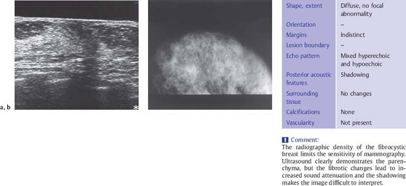
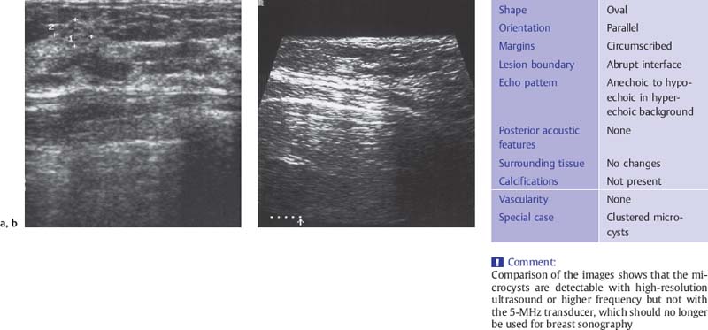
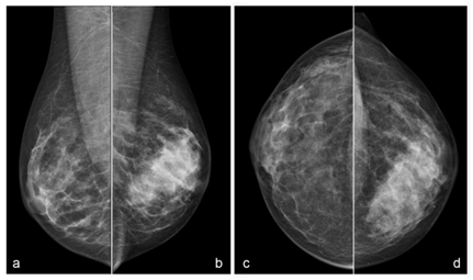
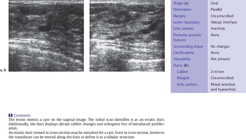
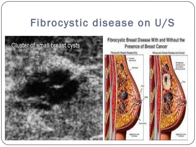
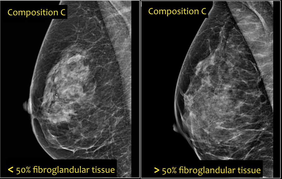
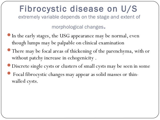
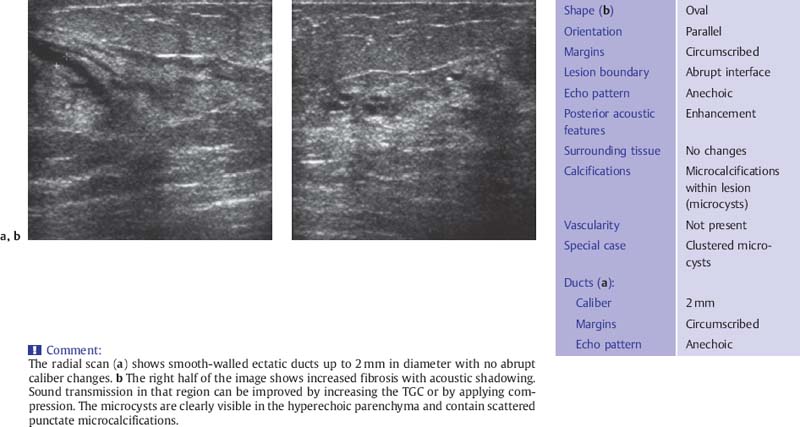
Fibrocystic breast disease ultrasound findings. It is not a disease but. Mammographic findings of fibrocystic change include a focal mass focal asymmetry microcalcifications architectural distortion areolar skin thickening and no abnormality 3031 whereas. The criteria for the ultrasound diagnosis of fibrocystic change are shown in Table 65.

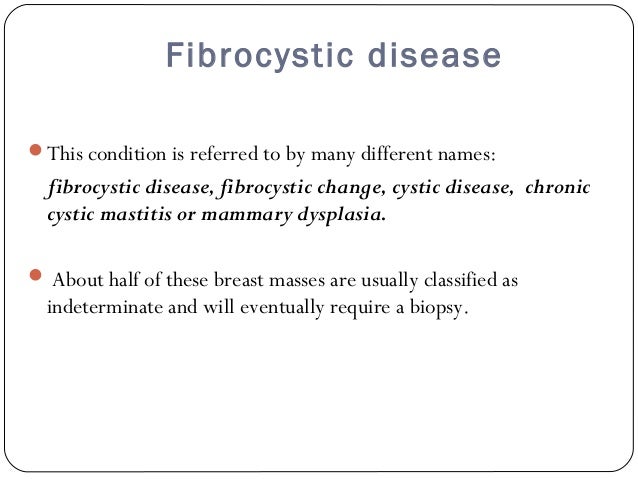
My primary clinical diagnosis is FIBRO-CYSTIC changes of the breast after correlating my physical examination findings with the ultrasound report of presence of subcentimeter solid and cystic nodules. Symptoms may worsen during certain parts of the menstrual cycle. The most common investigative tools to assess for these clinical findings are mammograms and ultrasound.
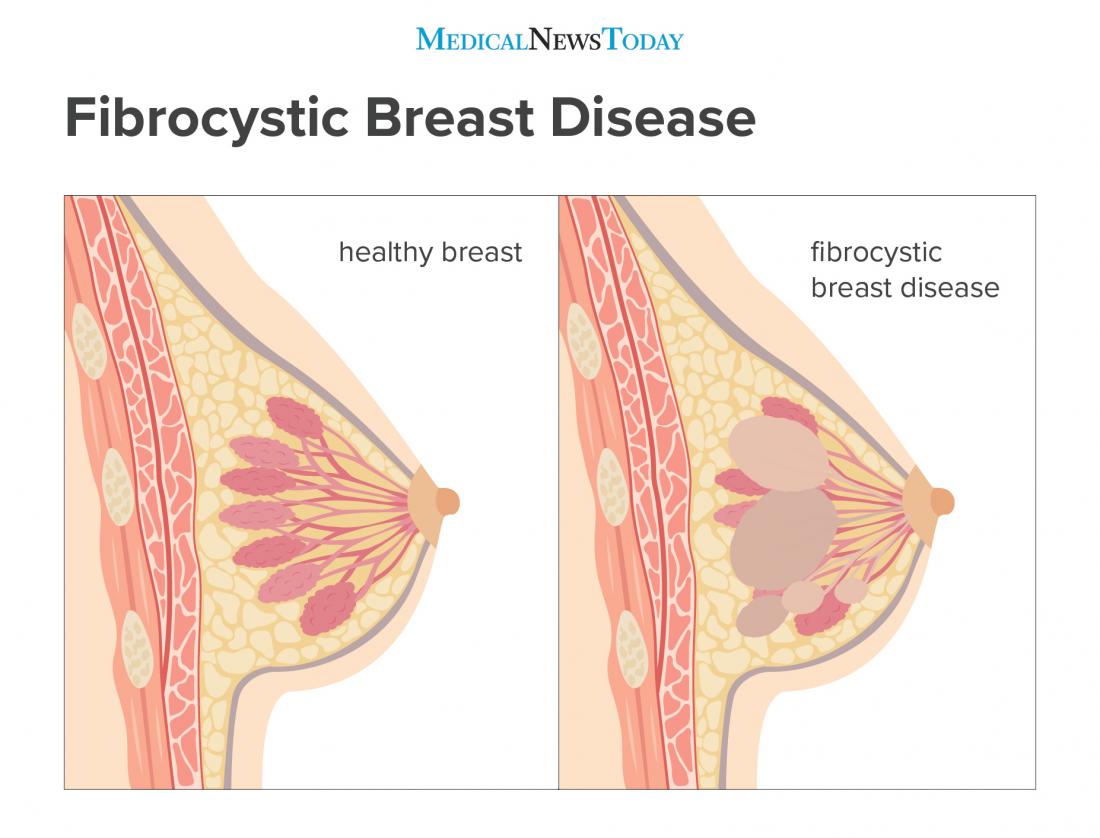
Fibrocystic breast changes is a condition of the breasts where there may be pain breast cysts and breast masses. It is the third most common breast lesion after fibrocystic disease and carcinoma. Some of them can even be irregular with shadowing often requiring a biopsy.

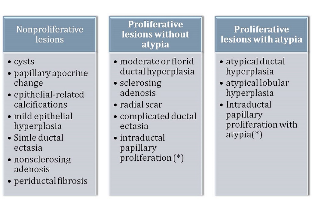
The clinical findings include symptoms such as dimpling of the skin peau deorange thickening pain and nipple discharge. The size is usually under 5 cm though larger fibroadenomas are known. Histologic findings that confirm fibrocystic breasts are apocrine metaplasia and hyperplasia gross and microscopic cysts and fibrosis.

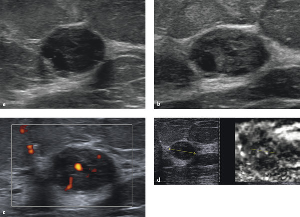
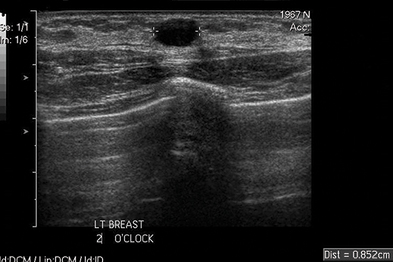
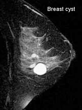
Sonographic findings in focal fibrocystic changes of the breast. 61 a b. There may be incidental findings of abnormalities on breast imaging mammography or breast ultrasound corresponding to breast cysts or a solid mass that on biopsy may confirm a diagnosis of fibrocystic breast.

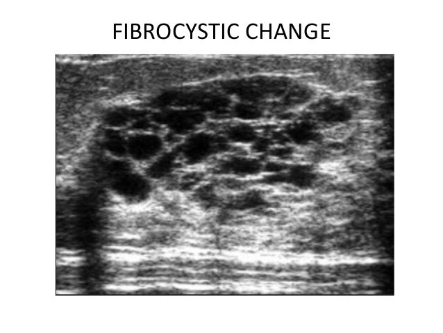
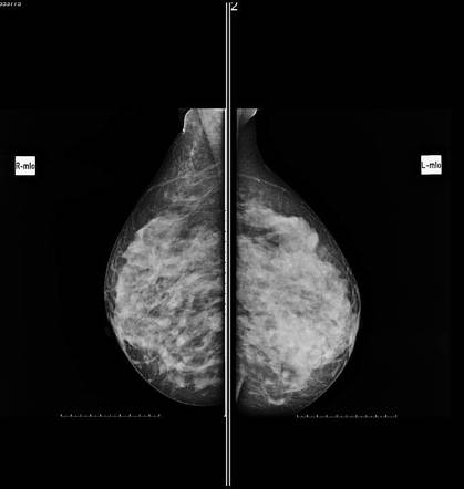
Researchers at Baylor College of Medicine and Womans Hospital of Texas studied 58 patients that had 60 lesions with a pathologic diagnosis of fibrocystic changes FC. Ultrasound findings vary from very echogenic breast tissue due to fibrosis to complex solid and cystic masses. Mammography may be used to differentially diagnose fibrocystic changes from breast cancer and other breast disorders.

On ultrasound findings may show. The breasts may be described as lumpy or doughy.

Fibrocystic Disease Breast Springerlink

Fibrocystic Disease Breast Springerlink

Ultrasound Characterization Of Breast Masses Abstract Europe Pmc

Ultrasound Characterization Of Breast Masses Abstract Europe Pmc

Us In A 40 Year Old Woman With Fibrocystic Disease Observers Arrived Download Scientific Diagram

Us In A 40 Year Old Woman With Fibrocystic Disease Observers Arrived Download Scientific Diagram

Fibrocystic Change Radiology Key

Fibrocystic Change Radiology Key

Us In A 47 Year Old Woman With Fibrocystic Disease The Mass Shows Six Download Scientific Diagram

Us In A 47 Year Old Woman With Fibrocystic Disease The Mass Shows Six Download Scientific Diagram

Ultrasound Characterization Of Breast Masses Abstract Europe Pmc

Ultrasound Characterization Of Breast Masses Abstract Europe Pmc

Fibrocystic Change Radiology Key

Fibrocystic Change Radiology Key

Radiology Medtube Net

Radiology Medtube Net

Ultrasound Characterization Of Breast Masses Abstract Europe Pmc

Ultrasound Characterization Of Breast Masses Abstract Europe Pmc

Fibrocystic Disease Of Breast Sono Scan 5d

Fibrocystic Disease Of Breast Sono Scan 5d

Something As Simple And As Complex As Fibrocystic Masthopathy Review Of Mri Findings In A Case Report Fernadez International Journal Of Radiology Radiation Therapy

Something As Simple And As Complex As Fibrocystic Masthopathy Review Of Mri Findings In A Case Report Fernadez International Journal Of Radiology Radiation Therapy

Breast Ultrasound Part 4 Fibrocystic Disease Of The Breast Youtube

Breast Ultrasound Part 4 Fibrocystic Disease Of The Breast Youtube

Radiological Features Of Breast Dx

Radiological Features Of Breast Dx

Figure 1 From Association Between Polycystic Ovary And Fibrocystic Breast Disease Semantic Scholar

Figure 1 From Association Between Polycystic Ovary And Fibrocystic Breast Disease Semantic Scholar

Fibrocystic Change Radiology Key

Fibrocystic Change Radiology Key

Breast Ultrasound Radiology Reference Article Radiopaedia Org

Breast Ultrasound Radiology Reference Article Radiopaedia Org

Epos C 1600

Epos C 1600

Fibrocystic Disease Breast Springerlink

Fibrocystic Disease Breast Springerlink

Clinical Cases Benign Lesions Radiology Key

Clinical Cases Benign Lesions Radiology Key

Plos One Ultrasound Guided Cable Free 13 Gauge Vacuum Assisted Biopsy Of Non Mass Breast Lesions

Plos One Ultrasound Guided Cable Free 13 Gauge Vacuum Assisted Biopsy Of Non Mass Breast Lesions

Breast Pathology Ultrasound

Breast Pathology Ultrasound

Focal Fibrocystic Changes Of The Breast As A Pseudotumoral Lesion Eurorad

Focal Fibrocystic Changes Of The Breast As A Pseudotumoral Lesion Eurorad

Inflammatory Breast Disease The Radiologist S Role Sciencedirect

Inflammatory Breast Disease The Radiologist S Role Sciencedirect

Https Encrypted Tbn0 Gstatic Com Images Q Tbn And9gcrisyvnpxqw3vjwoexrnd4fqm88ukaolrjxasmrbdyec4mxek7z Usqp Cau

Https Encrypted Tbn0 Gstatic Com Images Q Tbn And9gcrisyvnpxqw3vjwoexrnd4fqm88ukaolrjxasmrbdyec4mxek7z Usqp Cau

Ultrasound Breast Mass

Ultrasound Breast Mass

Fibrocystic Breast Disease Causes Symptoms Treatment

Fibrocystic Breast Disease Causes Symptoms Treatment

Breast Cyst Wikipedia

Breast Cyst Wikipedia

The Radiology Assistant Bi Rads For Mammography And Ultrasound 2013

The Radiology Assistant Bi Rads For Mammography And Ultrasound 2013

Clinical Review Benign Breast Disease Gponline

Clinical Review Benign Breast Disease Gponline

Mammogram Images Understanding Your Results

Mammogram Images Understanding Your Results

Fibrocystic Breast Disease Sonography Youtube

Fibrocystic Breast Disease Sonography Youtube

Multiple Fibroadenomas And Fibrocystic Disease Radiology Case Radiopaedia Org

Multiple Fibroadenomas And Fibrocystic Disease Radiology Case Radiopaedia Org

Secondary Infected Cysts A 35 Year Old Female With Fibrocystic Download Scientific Diagram

Secondary Infected Cysts A 35 Year Old Female With Fibrocystic Download Scientific Diagram

A 50 Year Old Woman With Fibrocystic Changes In The Left Breast Download Scientific Diagram

A 50 Year Old Woman With Fibrocystic Changes In The Left Breast Download Scientific Diagram

Ultrasound Breast Mass

Ultrasound Breast Mass

Health Care Provider Faqs Densebreast Info Inc

Health Care Provider Faqs Densebreast Info Inc

Ultrasound Characterization Of Breast Masses Abstract Europe Pmc

Ultrasound Characterization Of Breast Masses Abstract Europe Pmc

Epos C 1237

Epos C 1237

Ultrasound Breast Mass

Ultrasound Breast Mass

Fibrocystic Breast Disease Moose And Doc

Fibrocystic Breast Disease Moose And Doc

Fibrocystic Breast Disease Moose And Doc

Fibrocystic Breast Disease Moose And Doc

Faq S At Palm Beach Breast Institute By Dr Robert Gardner

Faq S At Palm Beach Breast Institute By Dr Robert Gardner

Fibrocystic Breast Changes Wikipedia

Fibrocystic Breast Changes Wikipedia

The Radiology Assistant Mri Of The Breast

The Radiology Assistant Mri Of The Breast

Fibrocystic Breast Disease Causes Symptoms And Treatment

Fibrocystic Breast Disease Causes Symptoms And Treatment

Juvenile Papillomatosis Of The Breast Eurorad

Juvenile Papillomatosis Of The Breast Eurorad

Fibroadenoma Breast Radiology Reference Article Radiopaedia Org

Fibroadenoma Breast Radiology Reference Article Radiopaedia Org

Cystic Breast Lesions Rinaldi 2010 Journal Of Ultrasound In Medicine Wiley Online Library

Cystic Breast Lesions Rinaldi 2010 Journal Of Ultrasound In Medicine Wiley Online Library

1

1

The purpose of this study was to identify the spectrum of sonographic appearances in histologically proven focal fibrocystic changes FC of the breast to enhance understanding of imaging findings in this commonly encountered benign condition of the breast.

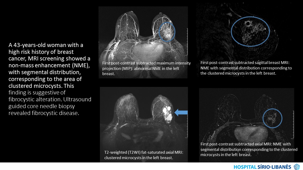
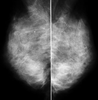
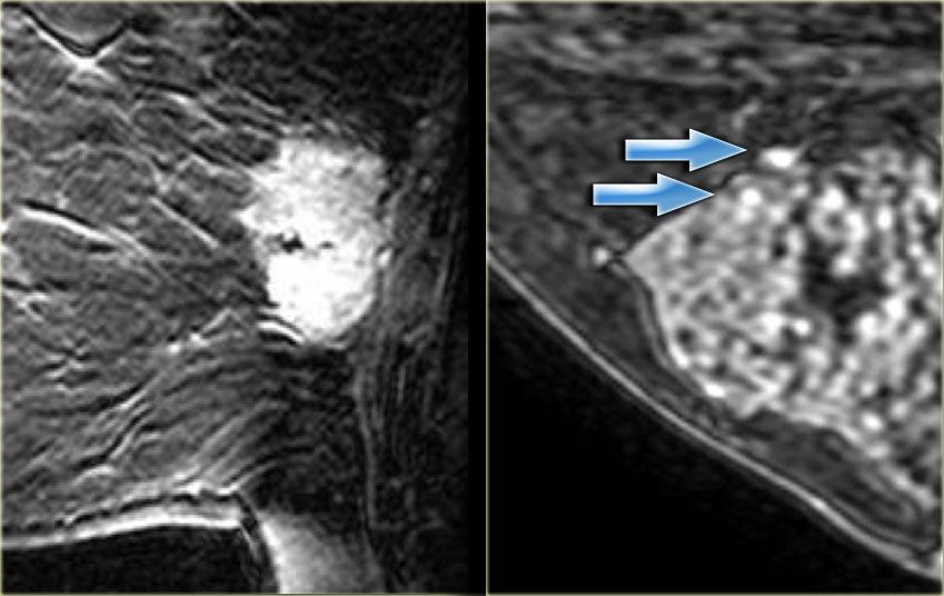
Fibrocystic breast changes is a condition of the breasts where there may be pain breast cysts and breast masses. Mammographic ultrasonographic and MR breast findings and anatomo-pathological analysis are described below. Ultrasound Review The sonographic findings in focal fibrocystic changes of the breast were described in a study recently published in Ultrasound Quarterly. It is rarely tender or painful. It usually presents as a firm smooth oval-shaped freely movable mass. The criteria for the ultrasound diagnosis of fibrocystic change are shown in Table 65. Fibrocystic breast changes is a condition of the breasts where there may be pain breast cysts and breast masses. Researchers at Baylor College of Medicine and Womans Hospital of Texas studied 58 patients that had 60 lesions with a pathologic diagnosis of fibrocystic changes FC. In the mammography we can see a left global asymmetry that primarily affects upper quadrants Figure 1 associating faint and few fine and heterogeneous scattered microcalcifications.


Fibrocystic breast changes can usually be diagnosed through a clinical breast exam and a symptom history. My primary clinical diagnosis is FIBRO-CYSTIC changes of the breast after correlating my physical examination findings with the ultrasound report of presence of subcentimeter solid and cystic nodules. Some of them can even be irregular with shadowing often requiring a biopsy. The size is usually under 5 cm though larger fibroadenomas are known. 61 a b. Your doctor also may use imaging tests such as mammography and ultrasound to examine the breasts. The most common investigative tools to assess for these clinical findings are mammograms and ultrasound.

Post a Comment for "Fibrocystic Breast Disease Ultrasound Findings"